

First Case of 2019 Novel Coronavirus in the United States | NEJM
On January 19, 2020, a 35-year-old man presented to an urgent care clinic in Snohomish County, Washington, with a 4-day history of cough and subjective fever. On checking into the clinic, the patient put on a mask in the waiting room. After waiting approximately 20 minutes, he was taken into an examination room and underwent evaluation by a provider. He disclosed that he had returned to Washington State on January 15 after traveling to visit family in Wuhan, China. The patient stated that he had seen a health alert from the U.S. Centers for Disease Control and Prevention (CDC) about the novel coronavirus outbreak in China and, because of his symptoms and recent travel, decided to see a health care provider.
Figure 1. Figure 1. Posteroanterior and Lateral Chest Radiographs, January 19, 2020 (Illness Day 4).
Apart from a history of hypertriglyceridemia, the patient was an otherwise healthy nonsmoker. The physical examination revealed a body temperature of 37.2°C, blood pressure of 134/87 mm Hg, pulse of 110 beats per minute, respiratory rate of 16 breaths per minute, and oxygen saturation of 96% while the patient was breathing ambient air. Lung auscultation revealed rhonchi, and chest radiography was performed, which was reported as showing no abnormalities (Figure 1). A rapid nucleic acid amplification test (NAAT) for influenza A and B was negative. A nasopharyngeal swab specimen was obtained and sent for detection of viral respiratory pathogens by NAAT; this was reported back within 48 hours as negative for all pathogens tested, including influenza A and B, parainfluenza, respiratory syncytial virus, rhinovirus, adenovirus, and four common coronavirus strains known to cause illness in humans (HKU1, NL63, 229E, and OC43).
Given the patient’s travel history, the local and state health departments were immediately notified. Together with the urgent care clinician, the Washington Department of Health notified the CDC Emergency Operations Center. Although the patient reported that he had not spent time at the Huanan seafood market and reported no known contact with ill persons during his travel to China, CDC staff concurred with the need to test the patient for 2019-nCoV on the basis of current CDC "persons under investigation" case definitions.8 Specimens were collected in accordance with CDC guidance and included serum and nasopharyngeal and oropharyngeal swab specimens. After specimen collection, the patient was discharged to home isolation with active monitoring by the local health department.
On January 20, 2020, the CDC confirmed that the patient’s nasopharyngeal and oropharyngeal swabs tested positive for 2019-nCoV by real-time reverse-transcriptase–polymerase-chain-reaction (rRT-PCR) assay. In coordination with CDC subject-matter experts, state and local health officials, emergency medical services, and hospital leadership and staff, the patient was admitted to an airborne-isolation unit at Providence Regional Medical Center for clinical observation, with health care workers following CDC recommendations for contact, droplet, and airborne precautions with eye protection.9
On admission, the patient reported persistent dry cough and a 2-day history of nausea and vomiting; he reported that he had no shortness of breath or chest pain. Vital signs were within normal ranges. On physical examination, the patient was found to have dry mucous membranes. The remainder of the examination was generally unremarkable. After admission, the patient received supportive care, including 2 liters of normal saline and ondansetron for nausea.
Figure 2.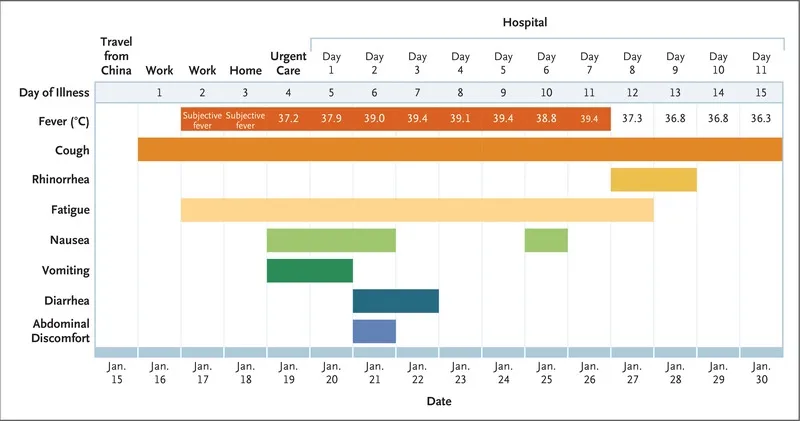
On days 2 through 5 of hospitalization (days 6 through 9 of illness), the patient’s vital signs remained largely stable, apart from the development of intermittent fevers accompanied by periods of tachycardia (Figure 2). The patient continued to report a nonproductive cough and appeared fatigued. On the afternoon of hospital day 2, the patient passed a loose bowel movement and reported abdominal discomfort. A second episode of loose stool was reported overnight; a sample of this stool was collected for rRT-PCR testing, along with additional respiratory specimens (nasopharyngeal and oropharyngeal) and serum. The stool and both respiratory specimens later tested positive by rRT-PCR for 2019-nCoV, whereas the serum remained negative.
Treatment during this time was largely supportive. For symptom management, the patient received, as needed, antipyretic therapy consisting of 650 mg of acetaminophen every 4 hours and 600 mg of ibuprofen every 6 hours. He also received 600 mg of guaifenesin for his continued cough and approximately 6 liters of normal saline over the first 6 days of hospitalization.
Table 1.
The nature of the patient isolation unit permitted only point-of-care laboratory testing initially; complete blood counts and serum chemical studies were available starting on hospital day 3. Laboratory results on hospital days 3 and 5 (illness days 7 and 9) reflected leukopenia, mild thrombocytopenia, and elevated levels of creatine kinase (Table 1). In addition, there were alterations in hepatic function measures: levels of alkaline phosphatase (68 U per liter), alanine aminotransferase (105 U per liter), aspartate aminotransferase (77 U per liter), and lactate dehydrogenase (465 U per liter) were all elevated on day 5 of hospitalization. Given the patient’s recurrent fevers, blood cultures were obtained on day 4; these have shown no growth to date.
Figure 3. Figure 3. Posteroanterior and Lateral Chest Radiographs, January 22, 2020 (Illness Day 7, Hospital Day 3).Figure 4.
A chest radiograph taken on hospital day 3 (illness day 7) was reported as showing no evidence of infiltrates or abnormalities (Figure 3). However, a second chest radiograph from the night of hospital day 5 (illness day 9) showed evidence of pneumonia in the lower lobe of the left lung (Figure 4). These radiographic findings coincided with a change in respiratory status starting on the evening of hospital day 5, when the patient’s oxygen saturation values as measured by pulse oximetry dropped to as low as 90% while he was breathing ambient air. On day 6, the patient was started on supplemental oxygen, delivered by nasal cannula at 2 liters per minute. Given the changing clinical presentation and concern about hospital-acquired pneumonia, treatment with vancomycin (a 1750-mg loading dose followed by 1 g administered intravenously every 8 hours) and cefepime (administered intravenously every 8 hours) was initiated.
Figure 5. Figure 5. Anteroposterior and Lateral Chest Radiographs, January 26, 2020 (Illness Day 10, Hospital Day 6).
On hospital day 6 (illness day 10), a fourth chest radiograph showed basilar streaky opacities in both lungs, a finding consistent with atypical pneumonia (Figure 5), and rales were noted in both lungs on auscultation. Given the radiographic findings, the decision to administer oxygen supplementation, the patient’s ongoing fevers, the persistent positive 2019-nCoV RNA at multiple sites, and published reports of the development of severe pneumonia3,4 at a period consistent with the development of radiographic pneumonia in this patient, clinicians pursued compassionate use of an investigational antiviral therapy. Treatment with intravenous remdesivir (a novel nucleotide analogue prodrug in development10,11) was initiated on the evening of day 7, and no adverse events were observed in association with the infusion. Vancomycin was discontinued on the evening of day 7, and cefepime was discontinued on the following day, after serial negative procalcitonin levels and negative nasal PCR testing for methicillin-resistant Staphylococcus aureus.
On hospital day 8 (illness day 12), the patient’s clinical condition improved. Supplemental oxygen was discontinued, and his oxygen saturation values improved to 94 to 96% while he was breathing ambient air. The previous bilateral lower-lobe rales were no longer present. His appetite improved, and he was asymptomatic aside from intermittent dry cough and rhinorrhea. As of January 30, 2020, the patient remains hospitalized. He is afebrile, and all symptoms have resolved with the exception of his cough, which is decreasing in severity.